Etude de cas : Douleur invalidante chronique du calcanéum, de l'astragal et de la partie proximal métatarsienne du pied dans un contexte post septicémique à Streptocoque A. Conséquences d’une anamnèse incomplète ou tardive. Relation ostéonécrose et septicémie.
Case study : Chronic and disabling pain of the calcaneum, astragal and proximal metatarsal of the foot in a context of a post septicemic episode (Streptococcus A). Consequences of a late or incomplete anamnesis. Link between osteonecrosis and septicemia.
Résumé :
Mme A. 62 ans, sans antécédents particuliers ni facteurs de risques sollicite dimanche 11 mars 2012 le médecin de garde pour profonde asthénie, suées nocturnes, frissons, douleurs abdominales et fièvre à 39°. Le médecin, devant ce tableau, évoque un simple syndrome grippal. Elle omet de signaler des pertes gynécologiques importantes depuis une semaine environ, attendant le retour de vacances de son gynécologue pour s’en charger. Son état empirant, elle est hospitalisée 2 jours plus tard par son médecin traitant pour un syndrome de réaction inflammatoire systémique (SRIS) avec abdomen défensif. Elle ressent une douleur intense sur le bord externe du pied gauche invalidante qu’elle ne signale pas dans un premier temps.
Les examens biologiques révèlent une septicémie à Streptocoque Pyogène A1 sensible aux ATB habituellement adaptés, d’entrée gynécologique traitée par nettoyage chirurgical (coelioscopie, pose de drains) et antibiotique IV (Amoxicilline / Ofloxacine) 10 j, relayés par 10j d’Amoxicilline Per Os.
Mme A. recouvre rapidement un état normalisé en 48h, mais son pied est œdémateux et douloureux lui interdisant tout appui. Un traitement pour arthrite microcristalline par colchicine est entrepris (sans amélioration notable). Mme A. réalise des examens complémentaires : radio, doppler (recherche d’une cause ischémique), scanner encéphalique (contrôle d’une légère dysarthrie à l’admission) en hospitalisation, scintigraphie et IRM en ambulatoire. Une algoneurodystrophie est évoquée. La scintigraphie montre la localisation de lésions tarsiennes et méta-tarsiennes, enfin l’IRM écarte la présence d’un foyer septique rémanent d’ostéomyélite. A J+ 20 , Mme A. est toujours gênée avec cependant une lente amélioration de son état fonctionnel, la douleur revenant toutefois dès qu’elle sollicite son pied.
Mme A. évoque alors sur le tard un surmenage de son pied avant l’épisode septicémique qui oriente définitivement le dernier diagnostic en date d’ostéonécrose d’installation antérieure à l’épisode septicémique, sans établir formellement la part lésionnelle proprement due à la septicémie.
Mme A. évoque alors sur le tard un surmenage de son pied avant l’épisode septicémique qui oriente définitivement le dernier diagnostic en date d’ostéonécrose d’installation antérieure à l’épisode septicémique, sans établir formellement la part lésionnelle proprement due à la septicémie.
Abstract :
On Friday, March 11th Miss A., 62 year old woman, without any prior medical history or risk factors asked for emergency medical support. Symptoms were a deep asthenia, night sweats, shivers, abdominal pain, and fever of 102°. The physician on duty believes it's the flu. During her visit, Miss A. forgets to mention that she’s been having gynecological discharge for about a week and that she was waiting for her gynecologist to return from vacation to take care of the problem. In the meantime, her health continued to degrade. Two days later, as she was showing signs of systemic inflammatory reaction and contractile abdominal response to palpation, she was hospitalized by her attending physician. In addition, she had severe pain in her left foot, but failed to mention it during the admission process.
On Friday, March 11th Miss A., 62 year old woman, without any prior medical history or risk factors asked for emergency medical support. Symptoms were a deep asthenia, night sweats, shivers, abdominal pain, and fever of 102°. The physician on duty believes it's the flu. During her visit, Miss A. forgets to mention that she’s been having gynecological discharge for about a week and that she was waiting for her gynecologist to return from vacation to take care of the problem. In the meantime, her health continued to degrade. Two days later, as she was showing signs of systemic inflammatory reaction and contractile abdominal response to palpation, she was hospitalized by her attending physician. In addition, she had severe pain in her left foot, but failed to mention it during the admission process.
Blood tests revealed a septicemia caused by streptoccus pyogene A1, without resistance to antibiotics, most likely of gynecological origin. The peritonitis was surgically cleaned (coelioscopy followed by the installation of abdominal drains). Intravenous antibiotics amoxicilline and ofloxacine were given for 10 days, followed by amoxicilline per os for another 10 days.
Miss A. quickly returned to a normal state after 48h, but her left foot was still edematous and painful, causing complete disability of her left leg. Based on the observations of the hospital rheumatologist, he began treatment for microcristalline arthritis with colchicine, but this brought no noticeable improvement.
Miss A. then underwent additional tests which included x-ray, doppler ultrasound (research of ischemic cause of the lesion), encephalic TDM (control of dysarthrie noticed during the admission process), scintiscanning and MRI during ambulatory management. An algoneurodystrophy is evoked. The scintiscanning showed lesions of the tarsal and metatarsal regions, and the MRI ruled out the presence of a remnant septic center of osteomyelitis. At D+20, Miss A. was still constrained in her mobility and her foot pain returned as soon as she made the effort to walk. However, she showed slight improvement in her functional state.
Miss A. then underwent additional tests which included x-ray, doppler ultrasound (research of ischemic cause of the lesion), encephalic TDM (control of dysarthrie noticed during the admission process), scintiscanning and MRI during ambulatory management. An algoneurodystrophy is evoked. The scintiscanning showed lesions of the tarsal and metatarsal regions, and the MRI ruled out the presence of a remnant septic center of osteomyelitis. At D+20, Miss A. was still constrained in her mobility and her foot pain returned as soon as she made the effort to walk. However, she showed slight improvement in her functional state.
Miss A. evokes then on late, unusual solicitation of her foot prior to the septicemic episode which definitively ruled out the diagnosis of osteomyelitis and confirmed the last diagnosis to-date of osteonecrosis prior to the septicemic episode, without formally establishing the lesional share properly due to this septicemia.
Historique :
Historical :
Avant hospitalisation :
Début du mois de mars : pertes gynécologiques inhabituellement abondantes non signalées par la patiente lors de la première anamnèse.
Début du mois de mars : pertes gynécologiques inhabituellement abondantes non signalées par la patiente lors de la première anamnèse.
Mme A. a sollicité son pied pour jouer avec son petit-fils les jours précédent l’épisode. En retraite depuis février, Mme A. avait doublé son exercice de marche quotidienne (2h au lieu d'1h).
Vendredi 9 mars : Produit charcutier (boudin) consommé au repas du soir. Diarrhées vomissements nocturnes.
Mme A. pense à une intoxication alimentaire.
AEG, fièvre frissons. Appel au médecin de garde dimanche 11/03/12. Diagnostic de syndrome grippal. Prescription antalgiques (DOLIPRANE).
L’AEG se poursuit. La patiente ne s’alimente plus, a soif de sucre. Elle appelle son médecin traitant qui la fait hospitaliser le 13/03/12 vers 13h.
Before Hospitalization :
Since the beginning of march: Miss A. had an abundance of dirty gynecological discharges not reported by the patient at the time of the first anamnesis.
Since the beginning of march: Miss A. had an abundance of dirty gynecological discharges not reported by the patient at the time of the first anamnesis.
Miss. A. overused her foot to play with her grandson few days prior the episode. Retired since February, Miss A. had been walking twice as much during her daily workout (2h instead of 1h).
Friday, March 9th : she ingested pork product with the evening meal. The same night she experienced diarrhea and vomiting. Miss. A. believed that she had food poisoning.
Alteration of general state, fever and chills. She called the doctor on call Sunday, March 11th. Diagnosed with the flu and prescribed a common analgesic, (DOLIPRANE).
General state of the patient worsened. The patient no longer ate, was thirsty for sugary products. Call was places to the attending physician who hospitalized Miss A. on March 13th.
Hospitalisation du 13/03/12 au 27/03/12 :
Examen clinique d’admission :
Sensibilité abdominale diffuse fébrile à 39° associé à des vomissements.
Examen biologique à l'admission :
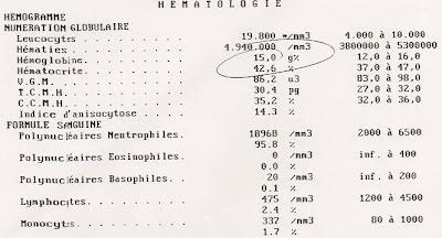
-syndrome infectieux inflammatoire : 19000 leucocytes, VS=104, CRP : 506(!!!)
-ECBU : urines stériles
-Hémocultures : streptocoque pyogène A sensible à la plupart des ATB testés.
Imagerie :
Le scanner abdomino-pelvien trouve un aspect de kyste hémorragique de l’ovaire droit, un épanchement du cul de sac de Douglas dont l’origine n’est pas évidente.
Radiographie du pied G normale.
Echo-Doppler artériel et veineux du membre inférieur gauche normal.
Prise en charge chirurgicale :
Dans le contexte de syndrome fébrile, avec pertes sales depuis quelques jours, une infection gynécologique est suspectée et la patiente est opérée le 14/03/12 en coelio chirurgie : constatation d’une pelvi-péritonite généralisée, nécessitant une toilette abdominale et péritonéale par lavage aux antiseptiques sans problème particulier, un drainage aspiratif de Redon, une antibiothérapie locale. Les prélèvements au niveau du cul de sac de Douglas s’avèrent négatifs.
Mme A. est placée sous AUGMENTIN en perfusion pendant 10 jours, 3g/j en association avec de l’Ofloxacine (OFLOCET). Puis AUGMENTIN PO 3g/j pendant 10j.
Evolution :
En 48h d’antibiothérapie, l’état de Mme A. est normalisé. L’épisode septicémique se termine donc favorablement. Cependant le pied gauche de Mme A. n’évolue pas (douloureux, oedemateux ) et reste donc préoccupant dans ce contexte post-septicémique.
Hospitalization from 13/03/2012 until 27/03/2012 :
Clinical examination during admission :
Feverish (102°) with a diffuse abdominal sensitivity associated with vomiting.
Biological examination :
Inflammatory infectious syndrome: 19000 leucocytes, VS=104, CRP: 506 (!!)
Inflammatory infectious syndrome: 19000 leucocytes, VS=104, CRP: 506 (!!)
- Cyto-Bacterial examination of urine : sterile
- Hemocultures: streptococcus pyogene A sensitive to the majority of the ATB tested.
Imagery :
The abdomino-pelvic scanner shows an aspect of hemorrhagic cyst of the right ovary, an outpouring of the bottom of the Douglas’pouch whose origin is not clear.
Radiography of the left foot is normal.
Arterial and venous Echo-Doppler of the left lower extremity is normal.
Surgical handling :
In the context of flu-like syndrome, with dirty discharge for several days, a gynecological infection was suspectded. The patient recieved surgery the 14th of March performing a coelioscopy that lead to following gestures:
- - Observation of a generalized pelvic-peritonitis.
- - Abdominal and peritoneal toilet with antiseptics without any special difficulty.
- - Drainage of Redon and local antibiotherapy.
- - Fluid cultures from the Douglas’pouch were negative.
Antibiotherapy :
Miss A. received 3g of AUGMENTIN IV a day for 10 days, with Ofloxacine for 7 days. Then AUGMENTIN per os for 10 days.
Evolution :
After 48 h of antibiotherapy, Miss A.‘s physical state is back to normal. Septicemic episode had a favorable outcome. However, her left foot still shows sign of inflammation, therefore still concerning in this context.
Examens conduits en ambulatoire suite à l'hospitalisation:
Scintigraphie :
Scintigraphy :
Scintigraphy :
CR Scintigraphie :
Lésions tarsiennes gauches intéressant surtout le calcanéum et l’astragale et, à un moindre degré, deux têtes métatarsiennes proximales.
Scintigraphy report :
Lesion of the left Tarsal, concerning mostly the dome of the astragal, and the distal part of the calcaneum as well as the 2nd and 5th head of the proximal metatarsal.
IRM :
IRM :
MRI :
CR IRM:
Petit foyer d'ostéonécrose du coin supéro-interne de l'astragale de 11mm. L'oedème osseux spongieux de la base du calcanéum et de la base du 5e métatarsien sont aspécifiques et ne paraissent pas de nature infectieuse (aucune alvéolyse et aucune réaction des parties molles de voisinage).
MRI report :
Concerning is the small area, about 11mm long, of osteonecrosis in the upper-internal astragal, of 11mm long. Cancellated bone edema concerning the basis of calcaneum and the base the 5th metatarsal are not specific and don’t seem to indicate an infectious nature (no alveolysis and no reaction of soft peripheral tissue).
Examens biologiques de contrôle :
NFS au 21/05/12
CBC on 05/21/12
CBC on 05/21/12
Normal excepté un niveau de vitamine D bas.
Normal except for a low level of D vitamin.
CONCLUSION :
Cette étude de cas illustre d’une part les conséquences d’une anamnèse incomplète : en l’occurrence Mme A. n’a pas signalé au premier médecin de garde ses pertes gynécologiques importantes les jours précédent sa fièvre, ce qui aurait naturellement pu orienter d’emblée le diagnostique vers une infection gynécologique invasive, au lieu d’une suspicion de syndrome grippal ce qui aurait permis une prise en charge plus précoce par ATB de la péritonite, et peut-être une atteinte moindre des os du tarse. (Cette hypothèse nécessiterait d' être approfondie sur des cas similaires).
Cette étude de cas illustre d’une part les conséquences d’une anamnèse incomplète : en l’occurrence Mme A. n’a pas signalé au premier médecin de garde ses pertes gynécologiques importantes les jours précédent sa fièvre, ce qui aurait naturellement pu orienter d’emblée le diagnostique vers une infection gynécologique invasive, au lieu d’une suspicion de syndrome grippal ce qui aurait permis une prise en charge plus précoce par ATB de la péritonite, et peut-être une atteinte moindre des os du tarse. (Cette hypothèse nécessiterait d' être approfondie sur des cas similaires).
La crainte de Mme A. au sortir de cet épisode septicémique douloureux, d’avoir un foyer infectieux rémanent a occulté dans sa mémoire la simple étiologie de surmenage de son pied (notamment lors de jeux avec son petits fils, et le doublement de sa ballade quotidienne, qu’ elle n’a évoqué que tardivement lors de la dernière visite au rhumatologue).L’orientation diagnostique définitive vers une ostéonécrose aux dépens d’une d’ostéomyélite ne s’est donc imposée que tardivement par le souvenir de cette sollicitation inhabituelle du pied ainsi que l’aspect à l’IRM des tissus environnant la lésion non évocateurs d’une infection.
D’autre part, le cas de Mme A. fournit un exemple de complication d’ostéonécrose de surmenage sur terrain ostéoporotique par un épisode septicémique. Il serait intéressant de voir dans quelle mesure le foyer d’ostéonécrose a été sur-lésé par le streptocoque A en particulier ou par le SRIS en général (atteinte ostéoblastes, déséquilibre calcique et carence en vitamine D pendant l’infection...), pour élargir la recherche à la prévalence de lésions d’ostéonécroses, dans des complications d’ostéomyélite chroniques, et par quels mécanismes une inflammation systémique aggrave de manière notoire une lésion ostéonécrotique prévalante.
CONCLUSION :
This study emphasizes the consequences of an incomplete anamnesis : Miss A. didn't
notify the first physician that she consulted regarding the gynecological discharge during the days prior to the fever. This information would have pointed towards a diagnosis of invasive gynecological infection, instead of the flu, which would have led to an earlier antibiotherapy of the peritonitis, and possibly a less severe lesion of the tarsal. (This assumption would need further study of similar cases).
Because of her painful experience with sever sepsis, Miss A. was concerned that she still might have a small area of infection in her foot. This anxiety made her forget about the simple etiology of overstraining her foot, specifically while she was playing with her grandchild, and doubling level of exercise twice. This was discovered during the last consultation with the rheumatologist). Diagnostic orientation towards osteonecrosis instead of osteomyelitis was reinforced more recently through, both, the memory of overstraining the foot and the aspect of peripheral tissue shown on the MRI not significant of an infection.
Furthermore, Miss A.'s case provides a good example of complication of osteonecrosis caused by overstraining on osteoporotic ground by a septicemia. It would be interesting to see how the pre-existing lesion of the tarsal might have been affected by the Streptococcus A specifically, or by the systemic inflammatory process in general (decreased osteoblastic action, lowered vitamin D during infection...), in order to be able to extend the path of research on the prevalence of osteoporotic lesion prior to chronic osteomyelitis, and by which mechanism, systemic inflammatory response can affect significantly a prevalent osteoporotic lesion.
Théophane Sylvestre (Schmitzberger) 2012


